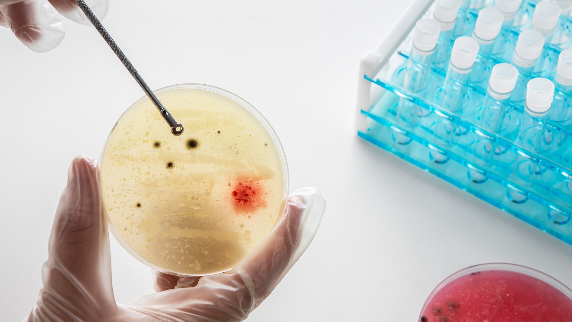
Total Body Checkup
Comprehensive organ and system diagnostics that detect early risks and reveal your body’s inner health — empowering prevention and longevity.
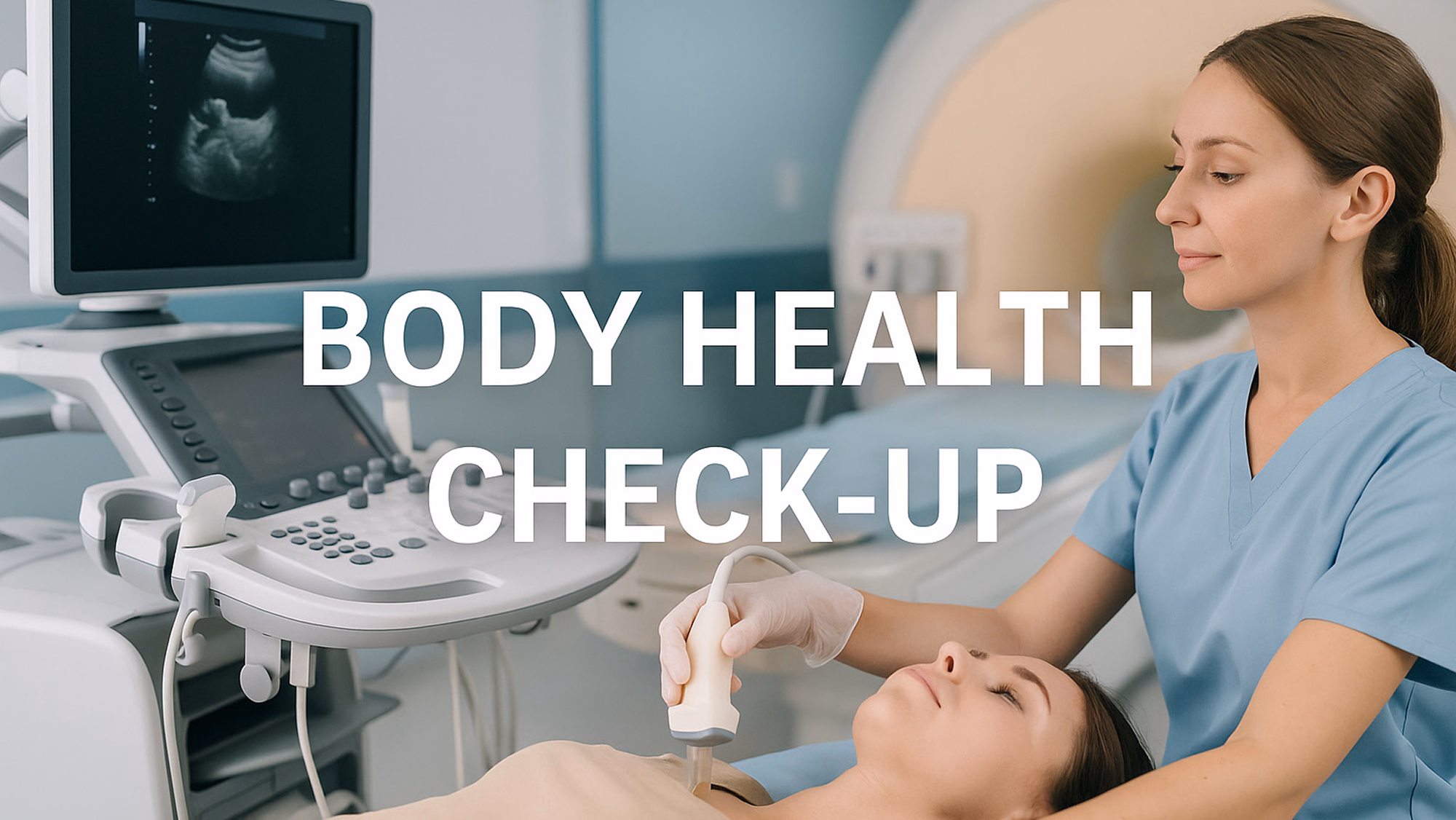
Total Body Health Checkup represents the next level of preventive medicine, a full-spectrum diagnostic program designed to evaluate vital organs, detect early signs of disease, and provide a clear picture of your internal health. This comprehensive assessment integrates 16 essential diagnostic programs, combining advanced imaging and organ-function analysis to uncover hidden imbalances before symptoms appear.
Among these, 13 programs are tailored for male health, focusing on the cardiovascular, liver, kidney, and metabolic systems, while 3 additional programs are designed exclusively for female health, covering breast and cervical screening. Female clients, however, can access all 16 programs for a complete and precise evaluation, ensuring both preventive and regenerative health optimization at every level.
This program comprises 16 specialized examinations designed to assess vital organs and systemic health, including:
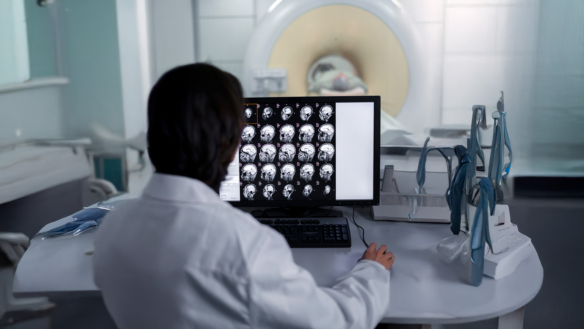
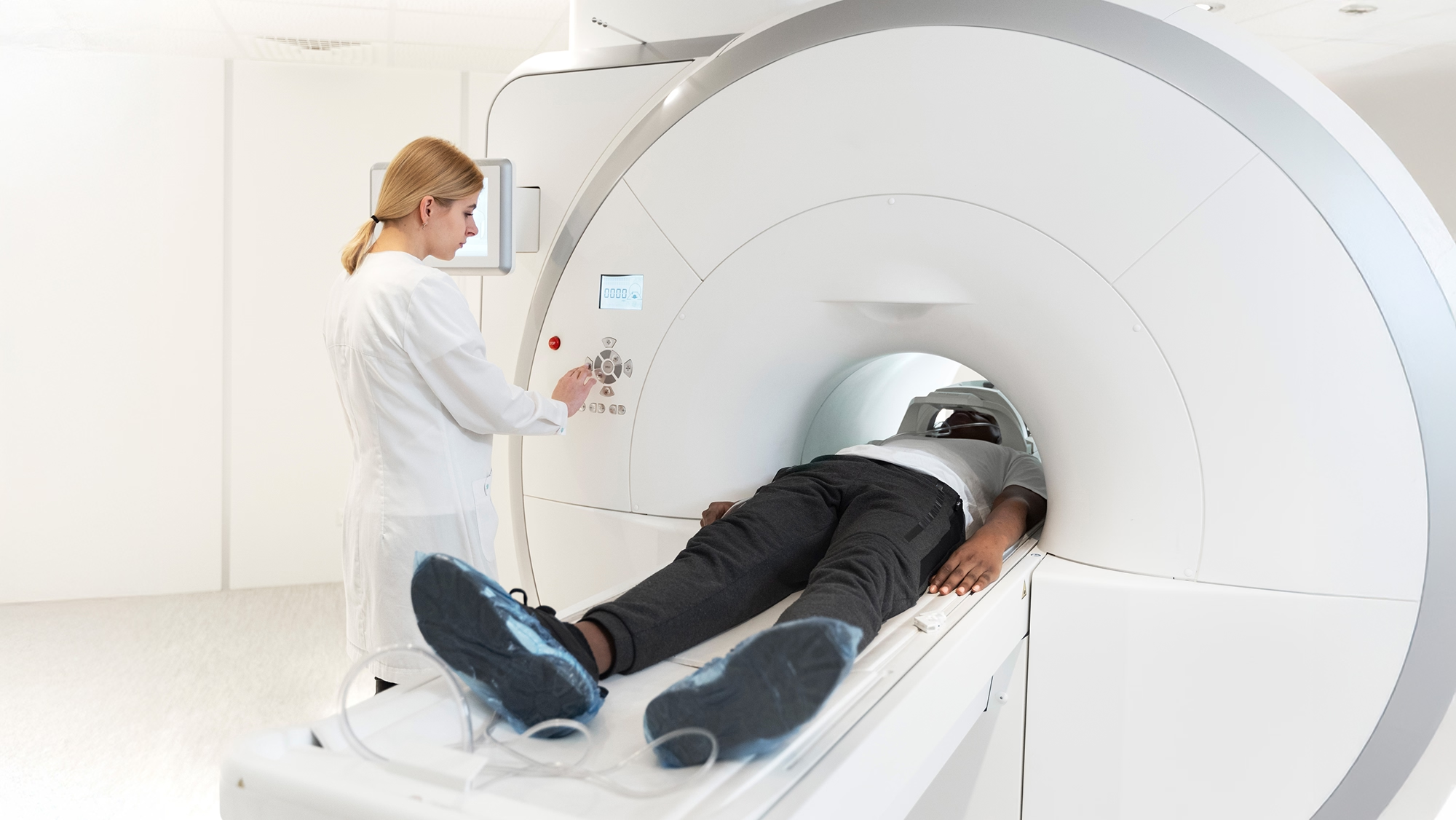
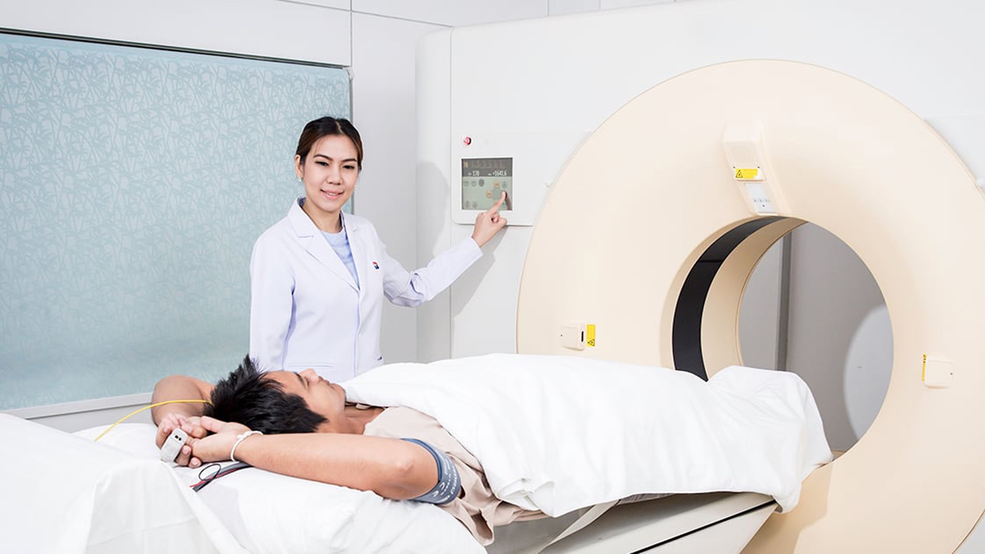
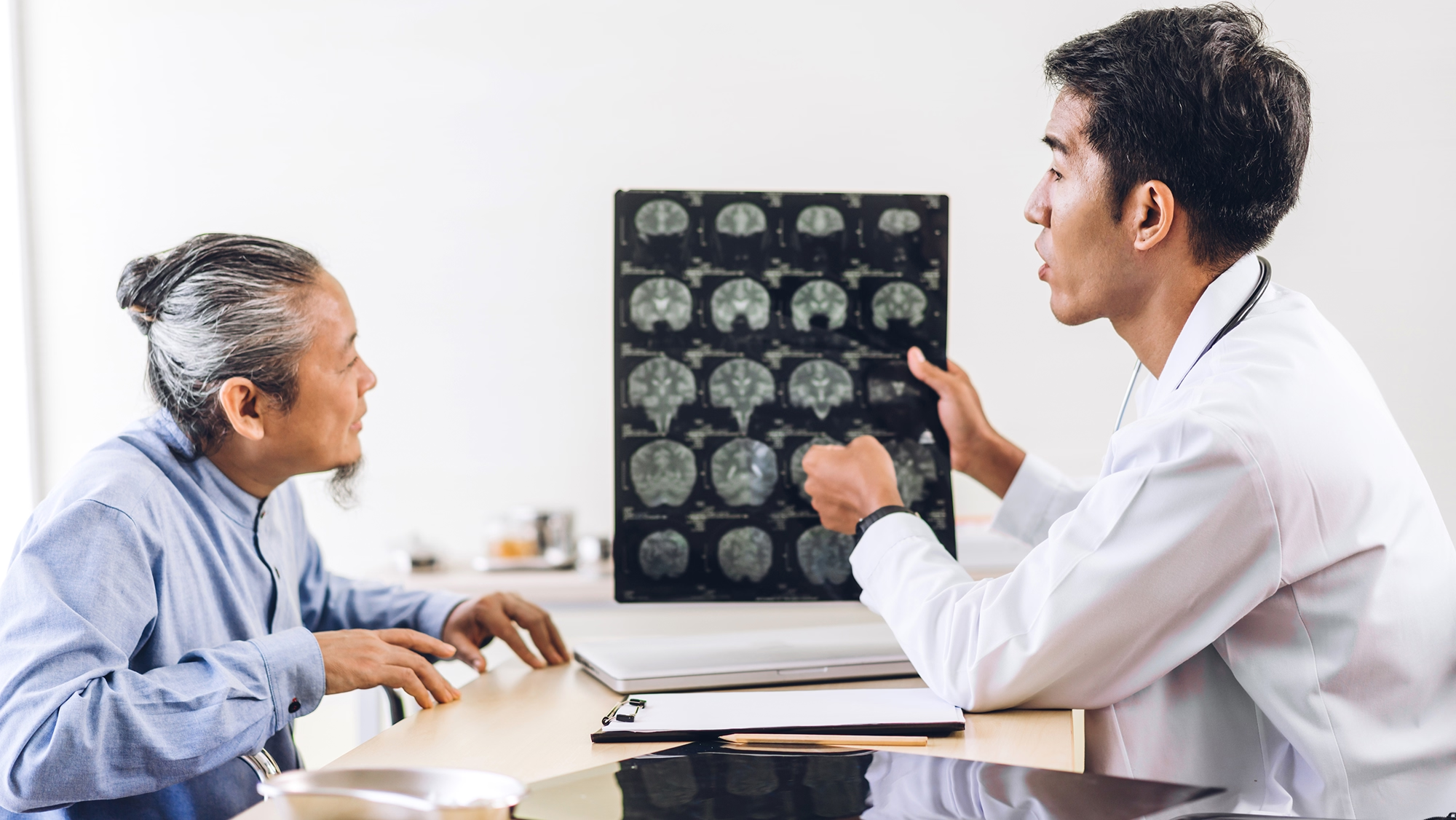
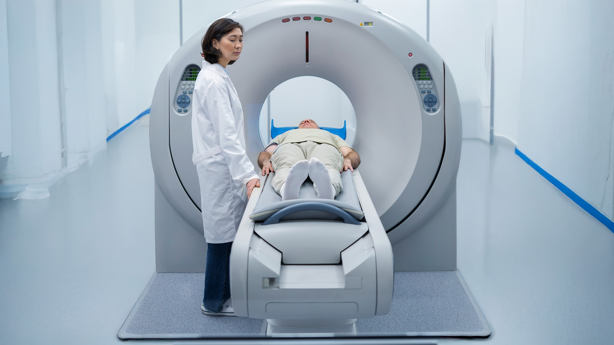
1. MRI Brain I+A+V
What is an MRI Brain I+A+V?
MRI Brain I+A+V is a highly detailed and comprehensive brain scan using MRI technology. It includes the use of intravenous contrast agents and specialized imaging of the brain’s venous system. This advanced diagnostic tool is particularly suitable for detecting complex neurological conditions or identifying potential abnormalities in the brain’s blood vessels.
MRI Brain I+A+V refers to a Magnetic Resonance Imaging (MRI) scan of the brain performed with the following parameters:
I = Intravenous contrast (usually gadolinium-based), which helps enhance certain structures or abnormalities such as tumors, infections, or inflammation.
A = Axial plane imaging, meaning the scan includes horizontal cross-sections of the brain.
V = Venography, which is imaging of the brain's venous system (veins), typically to evaluate for conditions like venous sinus thrombosis.
How MRI Brain I+A+V Works
MRI Brain I+A+V combines multiple imaging techniques into one powerful diagnostic scan to thoroughly assess the brain’s structure and blood vessels. Here's how it works step-by-step:
- You will be placed on a movable scanning table and instructed to remain as still as possible.
- A device containing coils is placed around your head to send and receive radio waves.
- An IV line will be placed in your arm to inject the contrast dye.
- The scanning table will be moved into the MRI machine, and the technologist will take multiple images.
- The entire procedure typically takes 45 to 60 minutes.
Key Benefits of MRI Brain I+A+V
- Non-invasive: The procedure does not involve radiation or surgery.
- Detailed Images: MRI provides highly detailed images of the brain and its structures.
- Diagnosis: Can help diagnose conditions like cerebral venous thrombosis and other venous abnormalities.
- Preoperative Imaging: Can be useful in preoperative imaging of brain tumors.
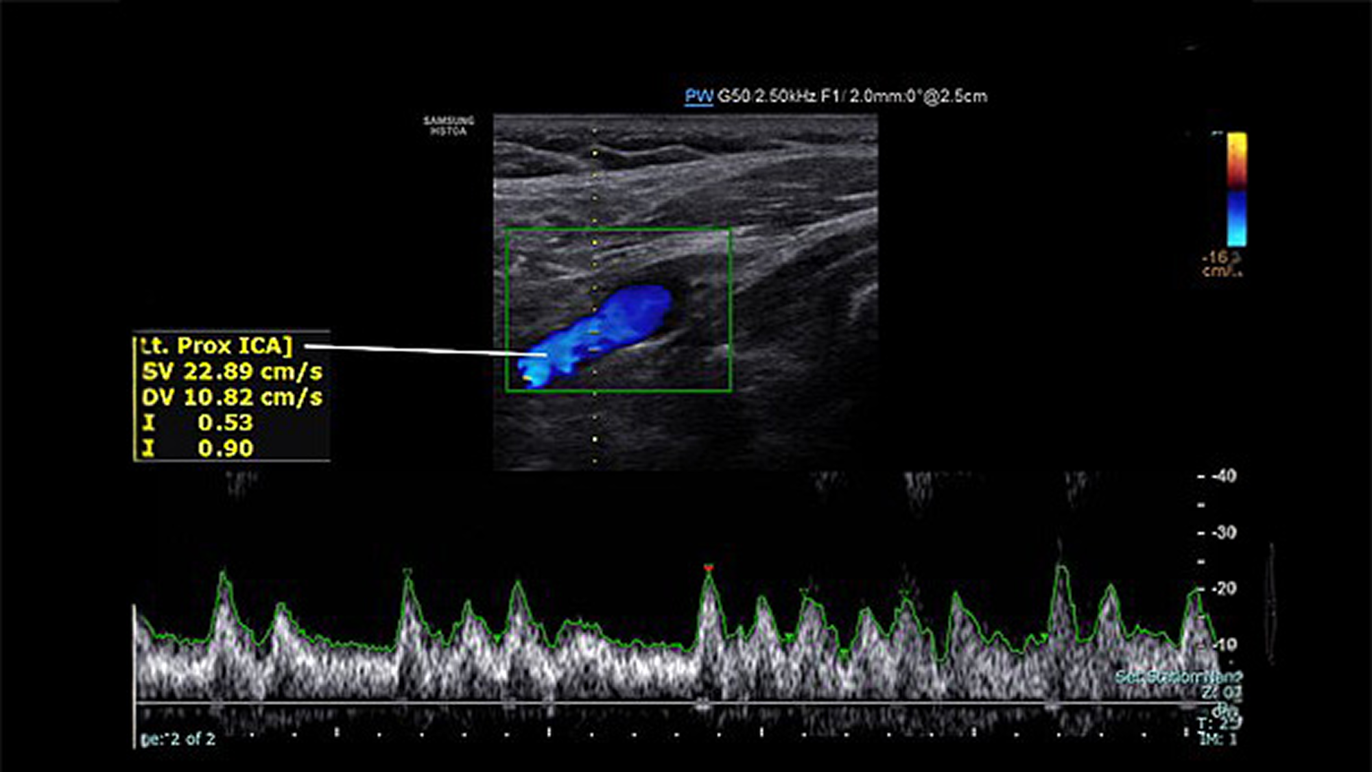
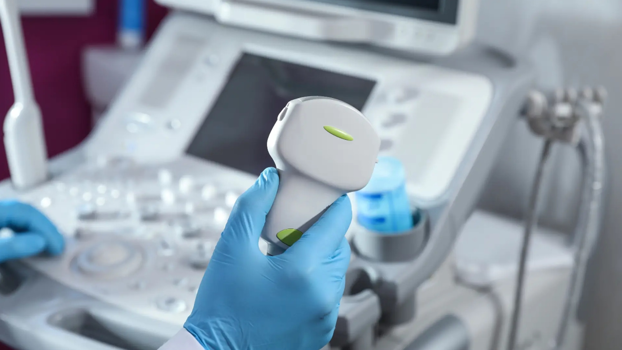
2. U/S Carotid
What is U/S Carotid?
A carotid ultrasound is a non-invasive imaging test that uses sound waves to examine the carotid arteries in the neck. These arteries are responsible for supplying blood to the brain, and the test helps detect blockages or narrowing in these arteries, which can increase the risk of stroke.

How U/S Carotid Works
- A gel is applied to the neck.
- A handheld device called a transducer is moved over the carotid arteries.
- High-frequency sound waves create real-time images of artery walls, blood flow patterns, and any irregularities like atherosclerosis (plaque buildup).
Key Benefits of U/S Carotid
- Non-invasive and safe.
- Early detection of vascular conditions.
- Real-time assessment of blood flow.
- Supports preventive care and risk stratification.
- Essential for surgical planning.
- Typically completed within 15 to 30 minutes and does not require any recovery time.
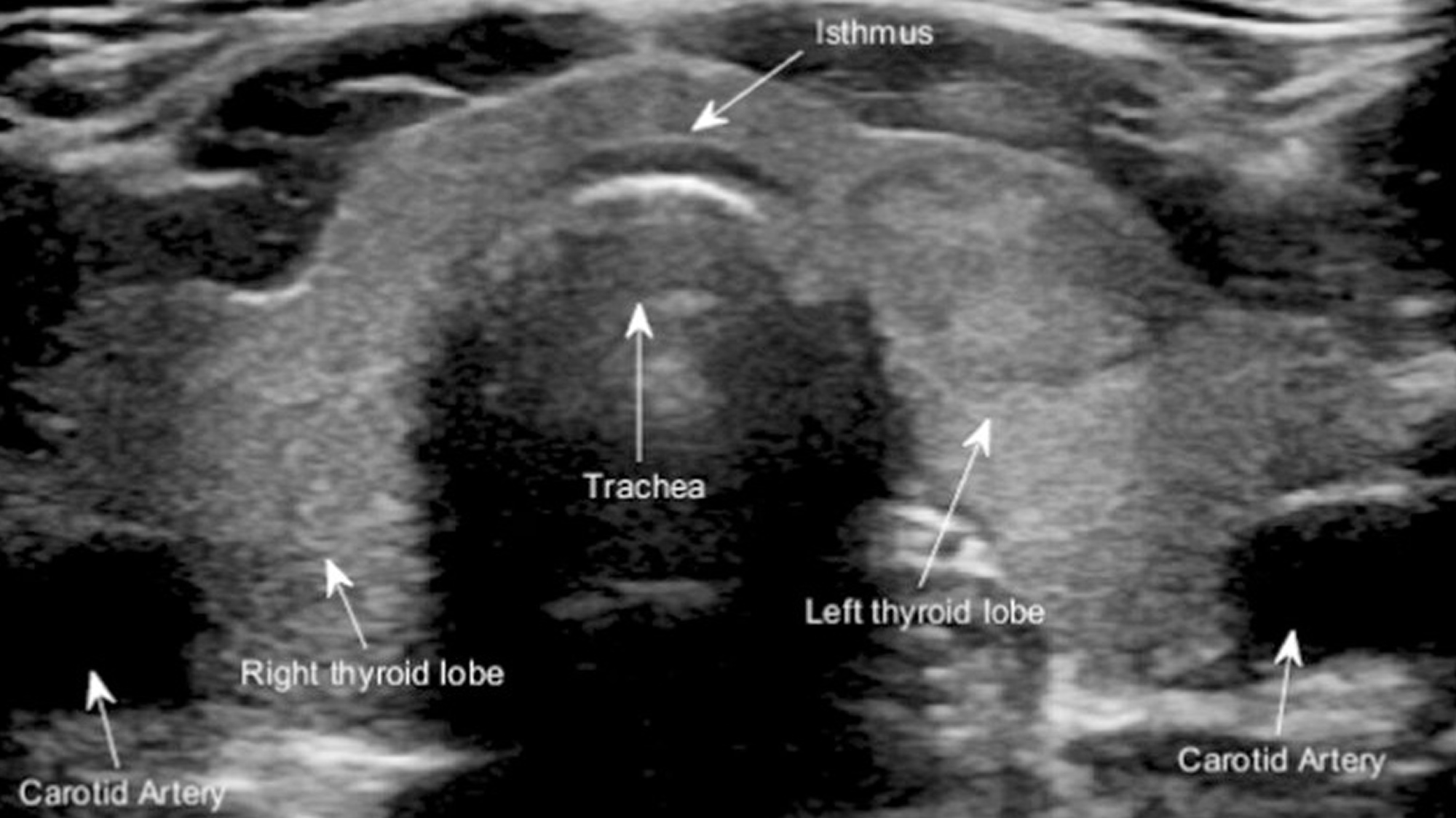
3. U/S Thyroid
What is U/S Thyroid?
A thyroid ultrasound is a painless imaging test that uses sound waves to create images of the thyroid gland and surrounding areas in the neck. It’s a common procedure used to diagnose thyroid conditions, assess the size and shape of the gland, and evaluate the presence of nodules or cysts.
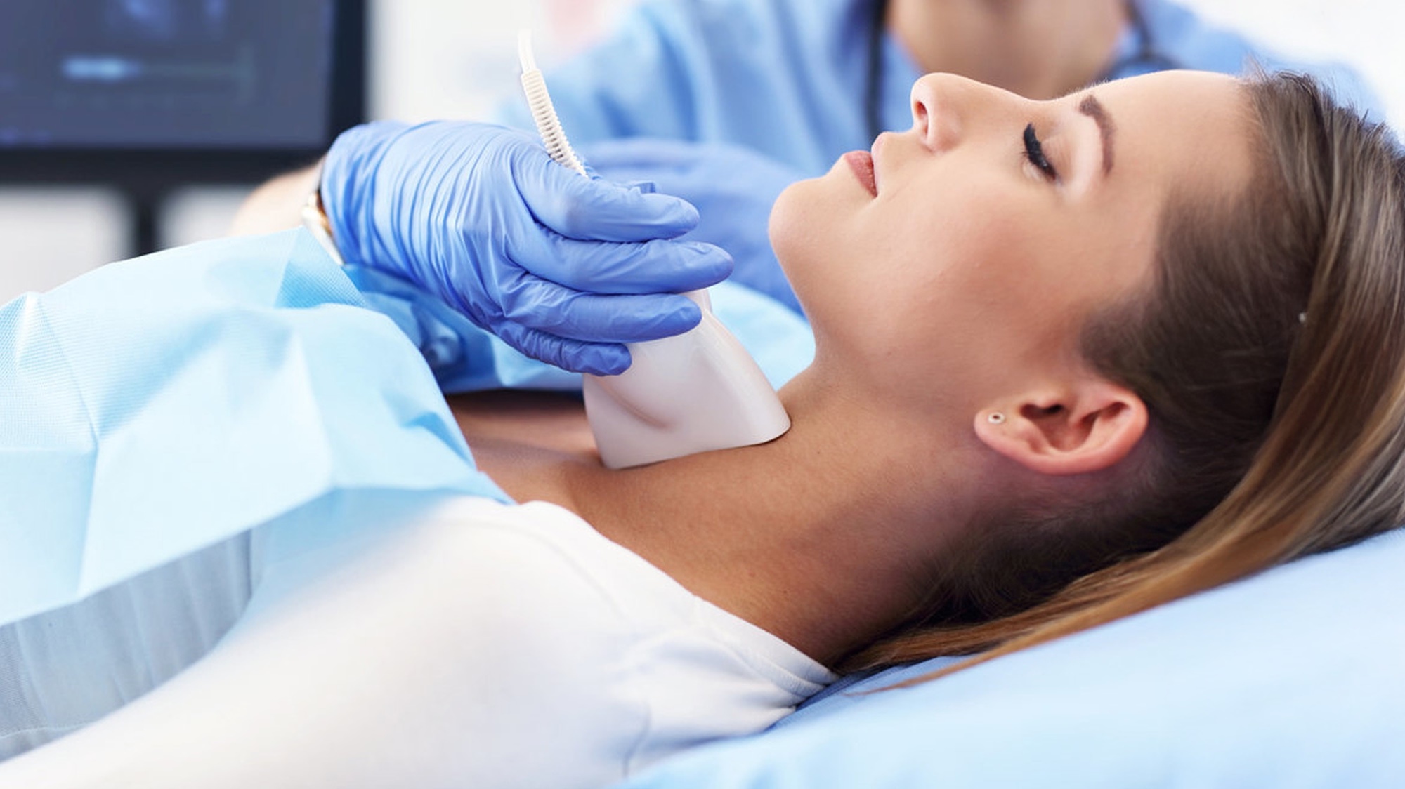
How U/S Thyroid Works
- Patients lie on their back with their neck extended.
- A water-based gel is applied to the skin on the neck to improve sound wave transmission.
- The transducer is moved over the thyroid, creating the images.
- The computer processes these echoes and displays live images on a monitor.
- A radiologist technician evaluates the shape, size, structure, and presence of any nodules or abnormalities.
- The procedure typically takes about 15–30 minutes.
Key Benefits of U/S Thyroid
- Non-Invasive & Painless: Completely safe for all ages, including pregnant individuals.
- No Radiation Exposure: Uses sound waves instead of ionizing radiation.
- Real-Time Imaging: Provides instant visualization of the thyroid gland and surrounding tissues.
- Highly Accurate Detection: Helps differentiate between benign and potentially malignant nodules.
- Early Diagnosis: Facilitates early detection of thyroid abnormalities, including thyroid cancer.
- Safe for Routine Monitoring: Ideal for follow-up in patients with known thyroid nodules or chronic thyroid conditions (e.g., Hashimoto’s thyroiditis).
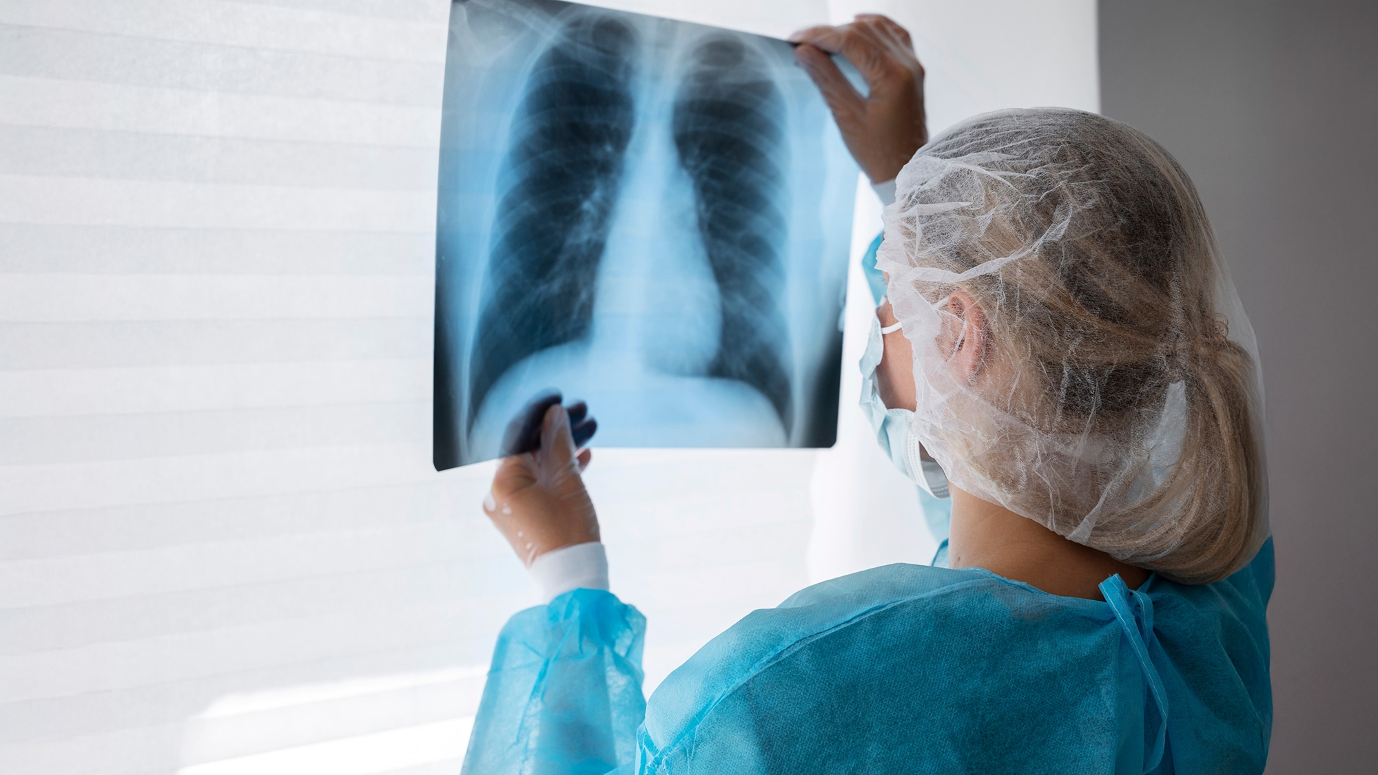
4. CT Chest Low Dose
What is CT Chest Low Dose?
CT Chest Low Dose is a non-invasive imaging test that uses X-rays and advanced computer technology to create detailed images of the lungs and chest, but with significantly reduced radiation exposure compared to standard CT scans. It is designed especially for routine screening rather than emergency diagnostics.
How CT Chest Low Dose Works
- Uses low-dose X-ray beams to capture high-resolution images.
- A spiral (helical) CT scanner rotates around the chest as the patient lies on a table.
- Produces cross-sectional slices that can detect small nodules, tumors, or inflammation.
- Entire procedure typically takes under 5 minutes, with minimal preparation.
Key Benefits of CT Chest Low Dose
- Finds cancer at an early, more treatable stage — often before symptoms appear.
- Uses up to 90% less radiation than a standard chest CT scan (~1.5 mSv vs. 7 mSv).
- Clinical studies show up to 20% reduction in lung cancer deaths when used for annual screening in high-risk groups.
- Safe for annual use, making it ideal for long-term monitoring in high-risk individuals.
- Accurate detection of lung abnormalities: identifies nodules, infections, scarring, emphysema, and other lung diseases.
- Safer for patients with kidney issues or allergies to contrast agents.
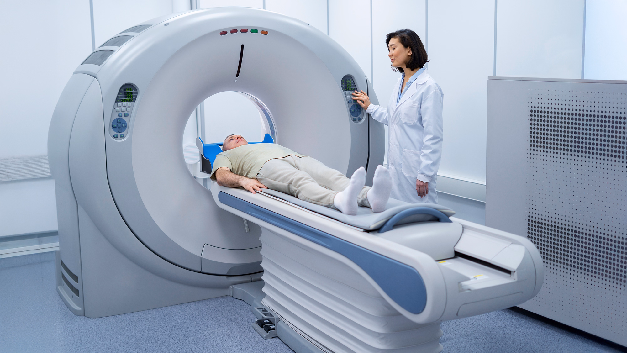
5. CT Calcium Score
What is CT Calcium Score?
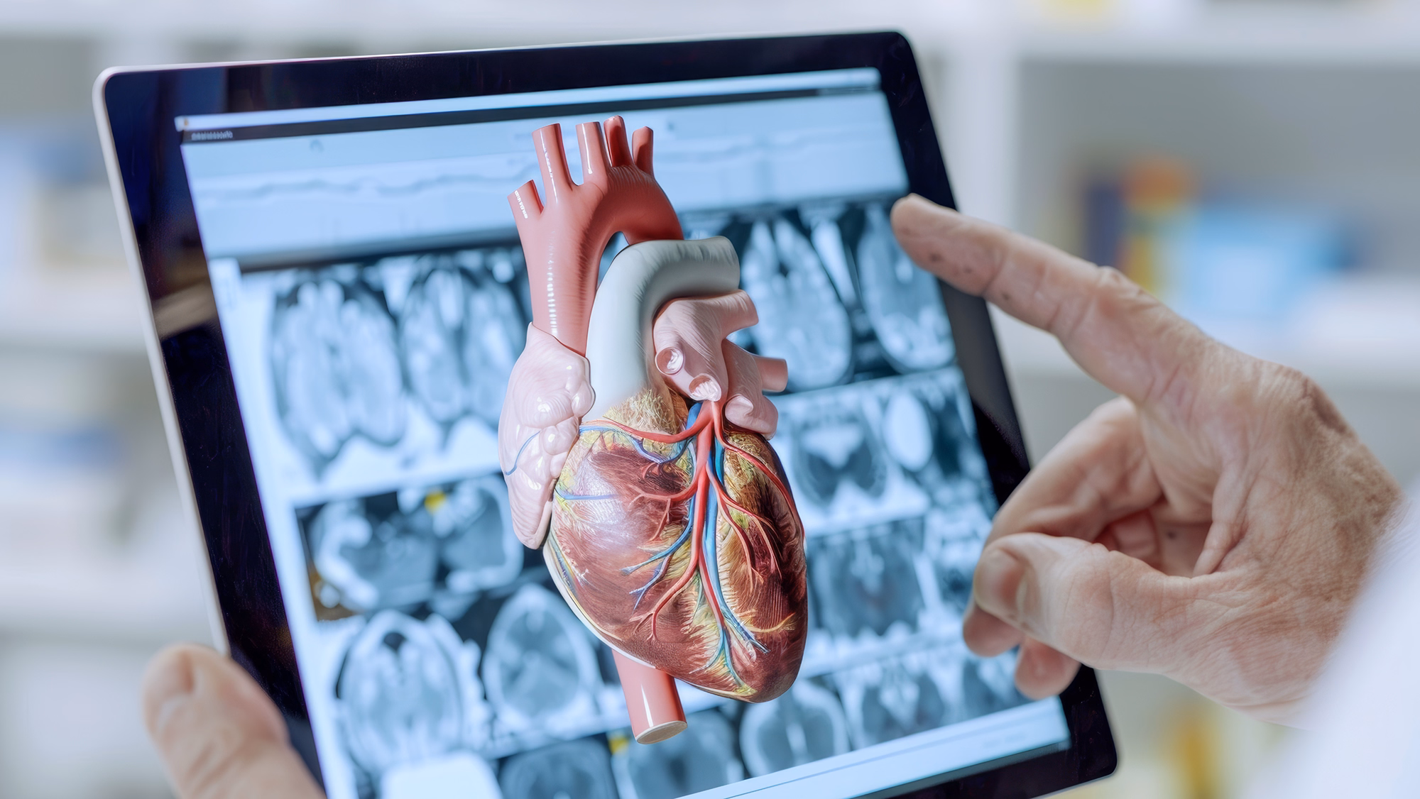
A CT Calcium Score, also known as Coronary Artery Calcium (CAC) Scoring, is a non-invasive diagnostic test that uses high-speed computed tomography (CT) to measure the amount of calcium deposits (calcified plaque) in the walls of the coronary arteries — the vessels that supply oxygen-rich blood to the heart.Calcium buildup in the arteries is a key indicator of atherosclerosis, a condition that increases the risk of coronary artery disease (CAD) and other cardiovascular events.
How CT Calcium Score Works
- Patients are advised to avoid caffeine or tobacco prior to the scan, as these may affect heart rate.
- Electrodes are placed on the chest to record the electrocardiogram (ECG), which synchronizes the scan with the cardiac cycle.
- The patient lies supine on the scanning table and is instructed to remain still and follow simple breathing instructions during image acquisition.
- A non-contrast CT scan is performed using ECG-gating to capture images during specific phases of the cardiac cycle.
- The CT data is processed using specialized software that detects calcified lesions in the coronary arteries.
- Each lesion is assigned a value based on density (Hounsfield units) and area. The total score reflects the overall severity of coronary atherosclerosis.
Key Benefits of CT Calcium Score
- Identifies calcified plaque in coronary arteries before symptoms appear, allowing for earlier intervention and prevention.
- Provides a measurable score (Agatston Score) that categorizes cardiovascular risk (low, moderate, or high).
- Helps tailor individualized treatment plans based on actual coronary artery calcium burden rather than generalized risk models.
- A simple and non-invasive scan that does not require needles, contrast dye, or anesthesia.
- Unlike CT angiography or cardiac catheterization, this test uses no contrast agents, making it safe for individuals with contrast allergies or kidney issues.
- Helps guide long-term lifestyle changes, follow-up schedules, and the use of medications to reduce future cardiovascular risk.
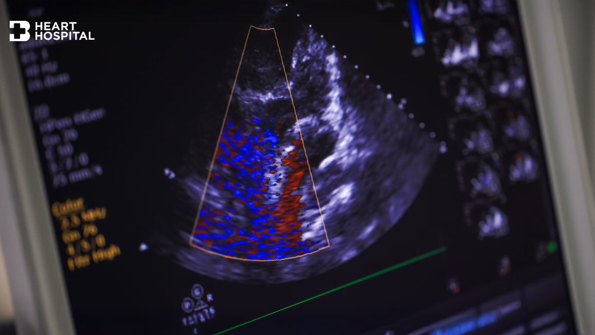
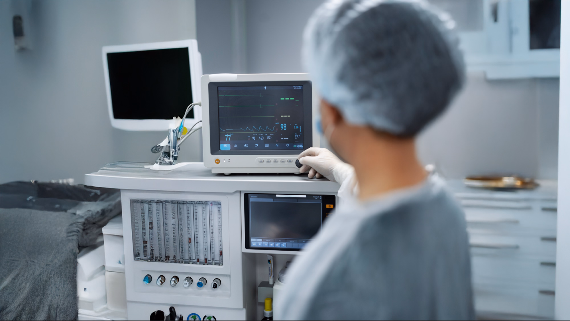
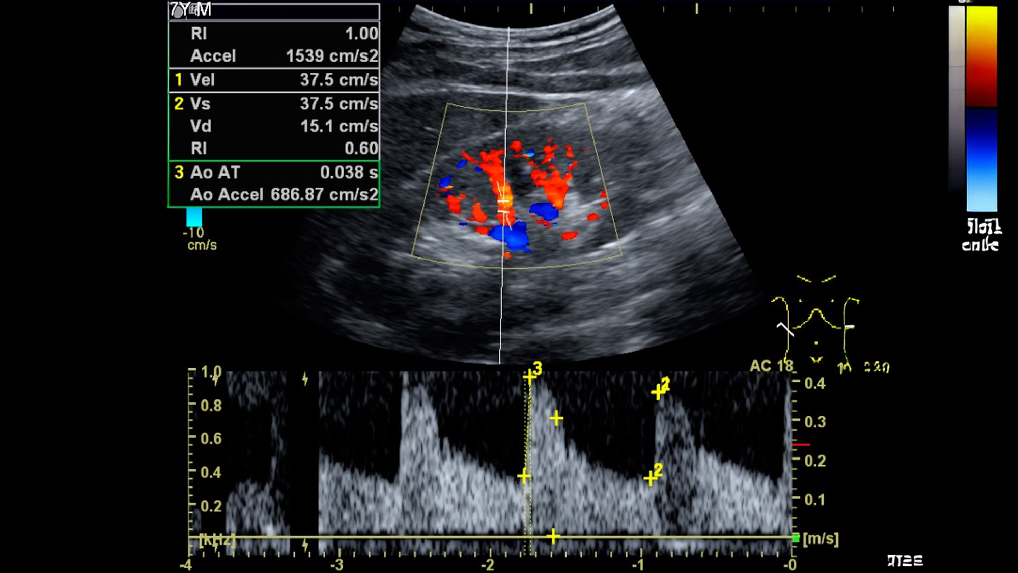
6. Echo Test (Echocardiogram)
What is Echo Test?
EKG or electrocardiography is a diagnostic test for the heart’s electrical activity. It is a quick, painless, and non-invasive method. Electrodes or sensors will be attached to your chest, arms, and legs while you lie down or exercise.
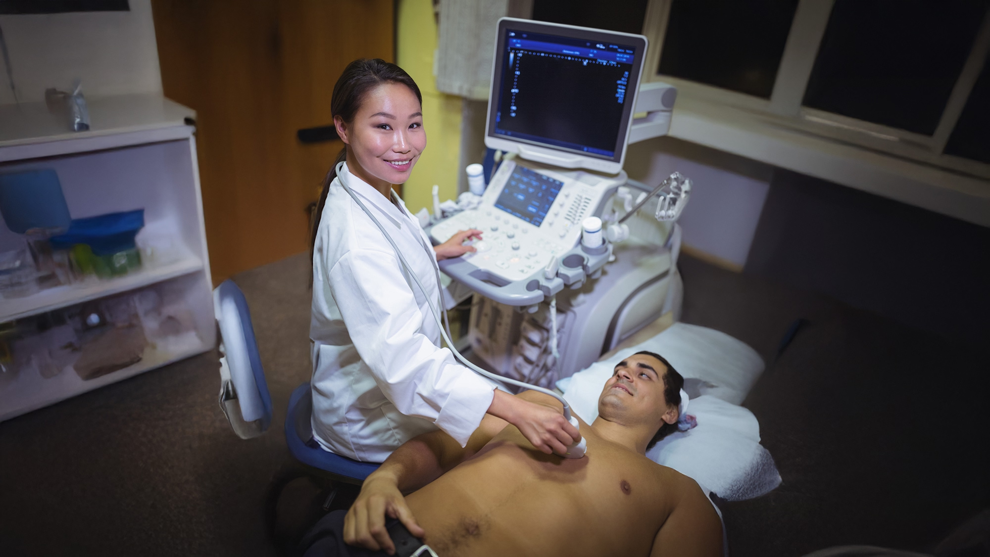
How Echo Test Works
- A technician places a small handheld device on your chest. This probe sends out sound waves.
- The sound waves bounce off the heart’s structures such as walls, chambers, and valves and return as echoes.
- The machine processes these echoes to create live images on a monitor. These images show: heart size and shape, movement of the heart walls, valve function, and blood flow patterns (using Doppler imaging).
Key Benefits of Echo Test
- Real-time visualization: Provides live images of the heart's structure and function.
- Early detection of heart conditions: Identifies abnormalities before symptoms appear.
- Monitors ongoing heart disease: Tracks progression and treatment response in conditions like heart failure or valve disease.
- Safe for all ages: Suitable for children, adults, and elderly patients.
- No radiation exposure: Uses ultrasound waves, making it safer than imaging tests with X-rays or CT scans.
- Quick and convenient: Often completed within 30–60 minutes with immediate preliminary results.
7. EKG (Electrocardiography)
What is EKG?
An echo test or echocardiogram is a non-invasive ultrasound test that provides real-time images of the heart. It helps evaluate the heart’s structure and function by using sound waves to create detailed pictures of the heart chambers, valves, and blood flow, including heart failure, valve problems, and structural abnormalities.
An EKG senses the electrical signal from the sinoatrial node, the heart’s natural pacemaker, tracks the heart muscle contraction, and sends the data to a computer which displays the recording in a wave pattern.
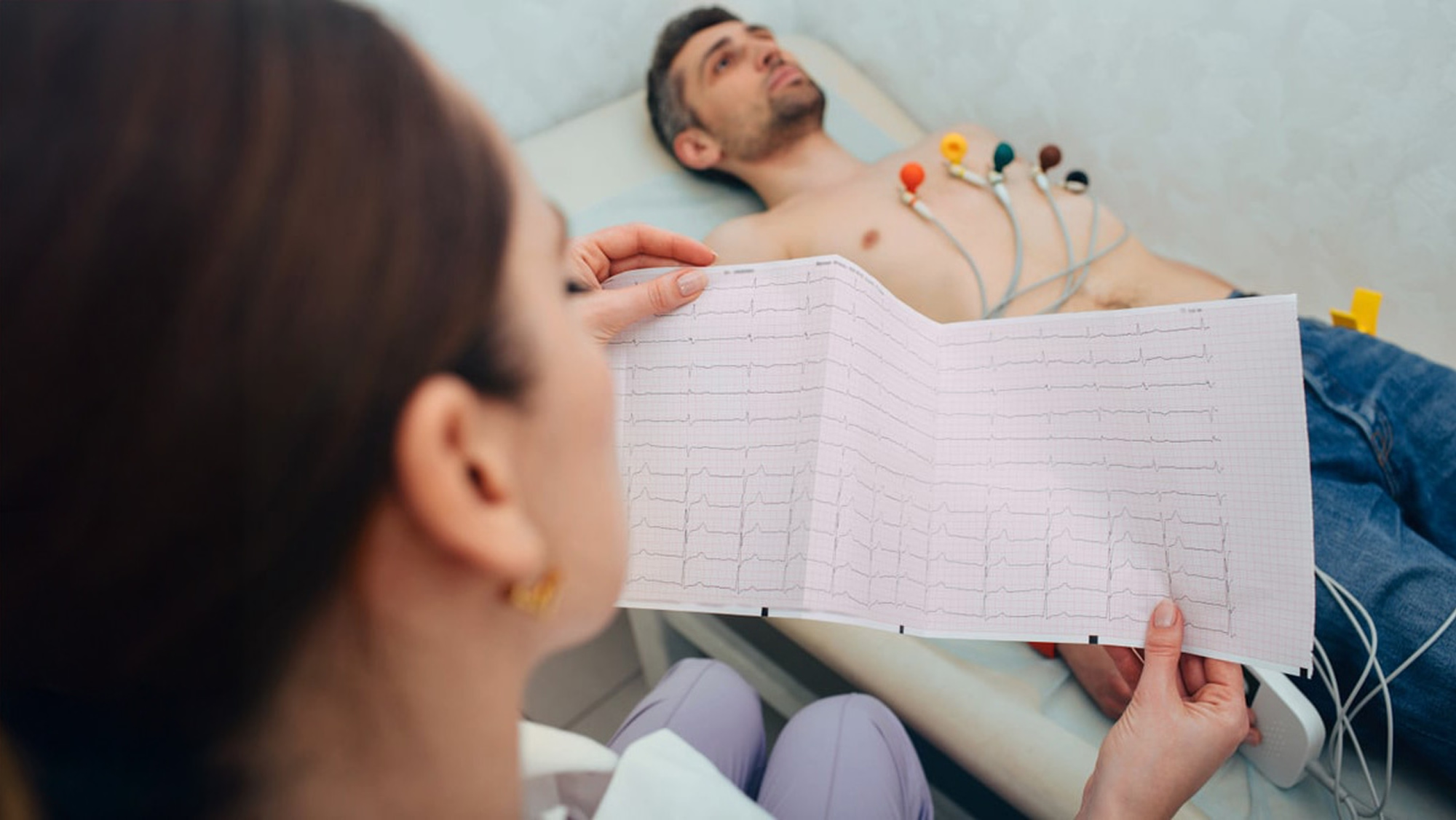
How EKG Works
- The patient is usually asked to lie down flat on a bed in a relaxed position.
- The chest, arms, and legs may be cleaned, and in some cases, small areas may be shaved to ensure proper electrode contact.
- During an EKG, 10 electrodes (small, sticky sensors) are placed on specific parts of the body — typically the chest, arms, and legs.
- The electrodes detect tiny electrical impulses generated with each heartbeat.
- The EKG machine records the strength and direction of the electrical signals and displays them as a waveform (graph) on a screen or prints it on paper.
Key Benefits of EKG
- Diagnosing heart problems: irregular heartbeats (arrhythmias), heart attack, chest pain, and other heart-related symptoms.
- Monitoring heart health: assessing pacemaker function, evaluating medication effectiveness, and monitoring before surgery.
- Screening for heart disease: family history, general population screening, individuals at low risk of heart disease.
- Early detection of heart conditions: identifies issues before symptoms arise, such as silent arrhythmias or early ischemia.
- Accessible and affordable: widely available in hospitals, clinics, and mobile health units.
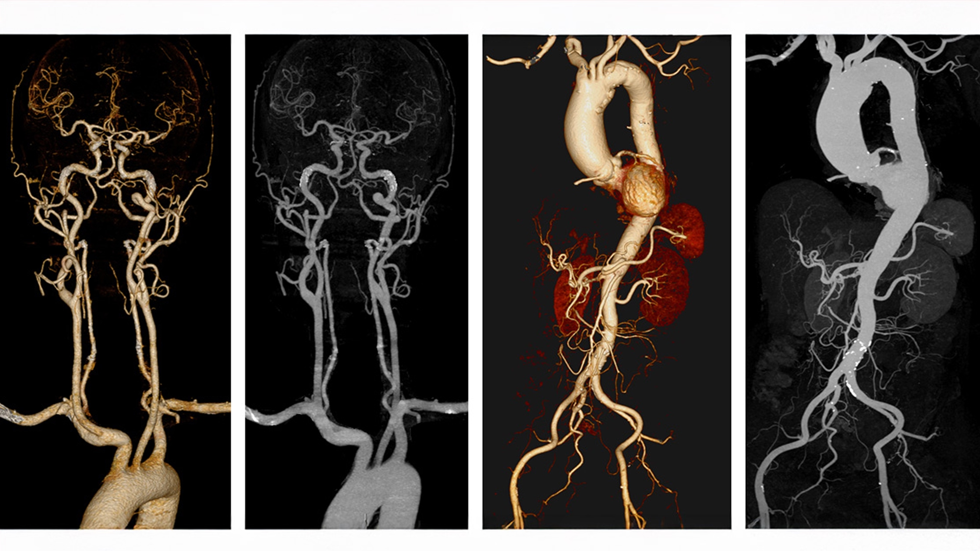
8. CTA with Contrast (Computed Tomography Angiography)
What is CTA with Contrast?
Computed Tomography Angiography (CTA) is an advanced imaging technique used to visualize blood vessels throughout the body. It primarily focuses on arteries in areas such as the neck and brain, coronary arteries, thoracic vessels, abdominal and pelvic vessels, and the arteries of the arms and legs.
During the procedure, a contrast agent (radiopaque dye) is injected intravenously through a pre-inserted needle, typically in the arm or leg. As the contrast circulates through the vascular system, the radiologic technologist performs high-resolution CT scanning using specialized techniques.
How CTA with Contrast Works
- The patient is positioned on a CT scanner table.
- An IV line is placed to allow the contrast dye to be injected into the bloodstream.
- The CT scanner takes a series of X-ray images as the contrast dye flows through the blood vessels.
- A computer processes these images to create detailed pictures of the blood vessels.
- A radiologist interprets the images to identify any abnormalities.
Key Benefits of CTA with Contrast
- High Spatial Resolution: Detects even small blockages or abnormalities.
- Whole-body evaluation: Can scan multiple vascular regions in one session.
- 3D Visualization: Enables better surgical planning and patient education.
- Emergency Use: Rapid evaluation in acute stroke, chest pain, or trauma.
- Post-operative Monitoring: Assess stents, grafts, and surgical repairs.
9. U/S Whole Abdomen
What is U/S Whole Abdomen?
A Whole Abdominal Ultrasound is a non-invasive diagnostic imaging procedure that uses high-frequency sound waves to generate real-time images of the internal organs within both the upper and lower abdominal cavity, as well as blood vessels within the abdomen. It is commonly used for both screening and diagnostic purposes and can be performed on both men and women.
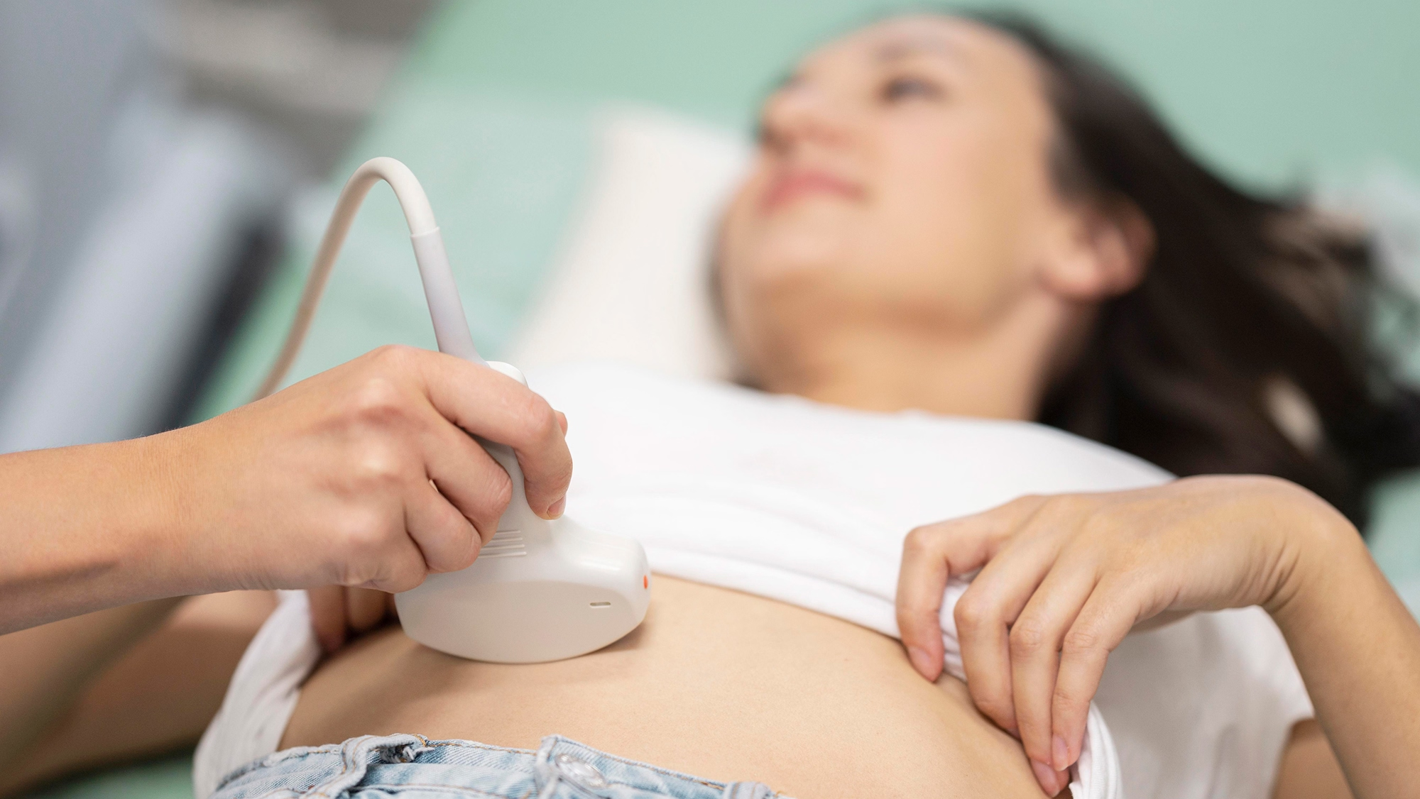
How U/S Whole Abdomen Works
- The patient lies on the examination table in a supine position.
- A water-based gel is applied to the abdomen to facilitate sound wave transmission.
- A transducer (probe) is moved across the abdomen to capture images.
- The reflected sound waves are converted into live images on a monitor for analysis.

Preparation Guidelines
- Fasting for 6–8 hours before the exam is recommended to minimize bowel gas, which may obscure organ visualization.
- For lower abdominal structures such as the bladder, patients may be asked to drink water to ensure a full bladder.
Organs Examined
- Liver: Evaluate for fatty liver, hepatomegaly, fibrosis, or tumors.
- Gallbladder: Detect gallstones, inflammation (cholecystitis), or biliary obstruction.
- Pancreas: Identify inflammation or masses.
- Kidneys: Look for kidney stones, cysts, or hydronephrosis.
- Spleen: Assess size and parenchymal abnormalities.
- Urinary Bladder: Detect residual urine volume, bladder wall abnormalities, or masses.
- Abdominal Aorta: Assess for aneurysms or vascular abnormalities.
- Prostate (male) / Uterus and Ovaries (female): As appropriate depending on patient’s sex.
Key Benefits of U/S Whole Abdomen
- Real-Time Dynamic Imaging: Allows visualization of organ movement and guides procedures such as fluid aspiration or biopsy.
- Early Detection of Pathologies: Enables identification of conditions such as liver diseases, gallbladder stones and inflammation, kidney cysts or stones, abdominal aortic aneurysms (AAA), and masses or tumors in abdominal organs.
- Monitoring of Chronic Conditions: Useful for tracking progression or regression of liver fibrosis, chronic kidney disease, and recurrent urinary tract issues.
- Supports Preventive Health Screening: Often used in routine check-ups for individuals with risk factors such as metabolic syndrome, hypertension, or family history of abdominal cancers.
10. Bone Density Scan (DEXA)
What is Bone Density Scan?
An echo test or echocardiogram is a nA Bone Density Scan, also known as DEXA (Dual-Energy X-ray Absorptiometry), is a gold-standard diagnostic test used to measure bone mineral density (BMD). It is highly accurate and widely utilized to diagnose osteopenia and osteoporosis, and to assess fracture risk.on-invasive ultrasound test that provides real-time images of the heart. It helps evaluate the heart’s structure and function by using sound waves to create detailed pictures of the heart chambers, valves, and blood flow, including heart failure, valve problems, and structural abnormalities.
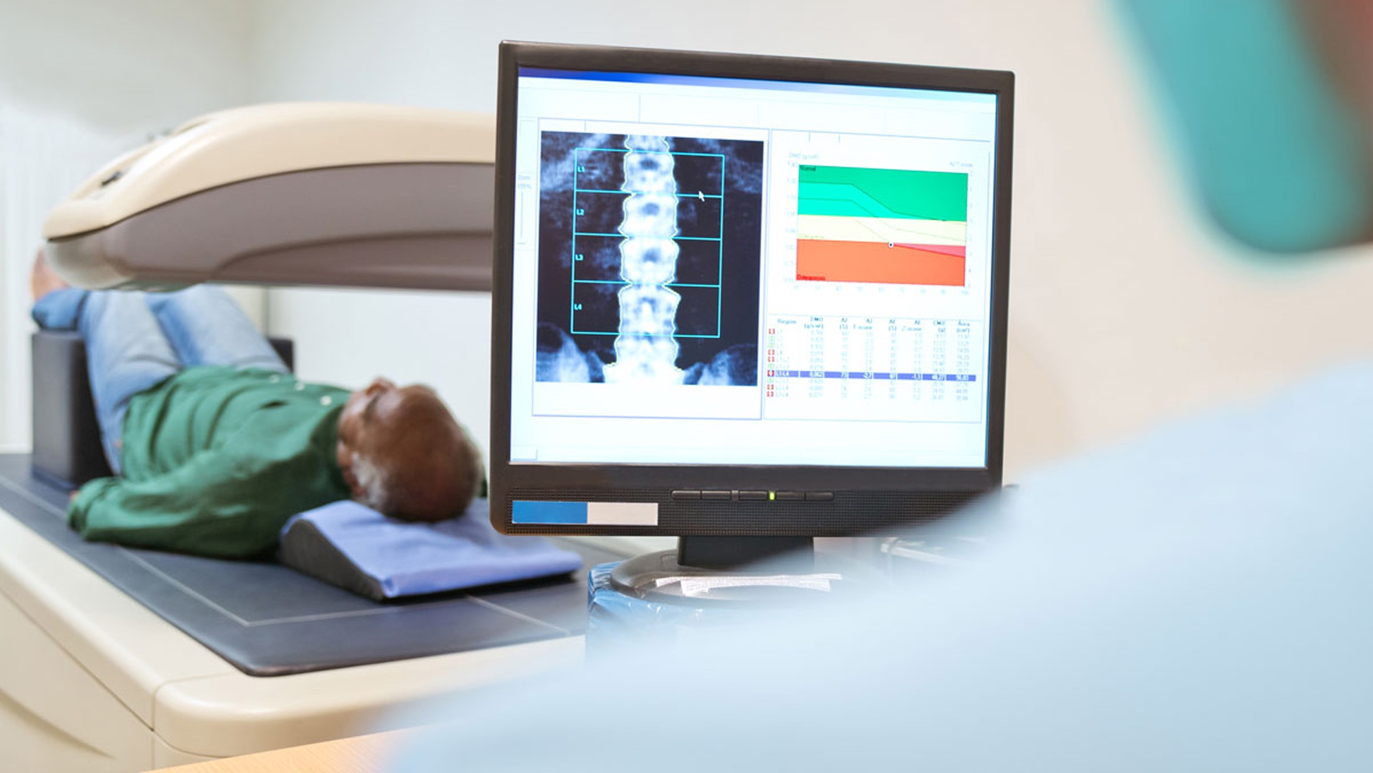
How Bone Density Scan Works
- The patient lies flat on a padded table, remaining still. The scan usually targets key sites of fracture risk:
Lumbar spine (L1–L4), Proximal femur (hip), Sometimes forearm (radius/ulna) in special cases. - The scanner emits two low-dose X-ray beams — one with high energy, one with low.
- A detector measures the amount of each beam that passes through the body. Because bone and soft tissue absorb X-rays differently, the machine can separate and measure just the bone component.
- Advanced software calculates the bone mineral content (grams of mineral per area) and generates a Bone Mineral Density (BMD) score, which is then compared to reference values:
T-score compares your BMD to that of a healthy young adult.
Z-score compares your BMD to someone of the same age, sex, and size.
Key Benefits of Bone Density Scan
- Accurate Fracture Risk Assessment: Helps predict risk of hip, spine, and wrist fractures.
- Gold-Standard Diagnostic Tool: Considered the most accurate and reliable method for assessing bone mineral density (BMD).
- Supports Management of Chronic Conditions: Detects bone loss in patients with long-term steroid use, menopause, cancer therapies, endocrine or autoimmune disorders.
- Crucial for At-Risk Populations: Recommended for postmenopausal women, elderly individuals, and people with a family history of fractures or osteoporosis.
- Aids in Lifestyle and Nutritional Planning: Calcium/vitamin D supplementation, diet and exercise plans, or fall prevention strategies.
11. Fibro Scan
What is Fibro Scan?
Fibro Scan, also known as Transient Elastography, is an advanced, non-invasive imaging modality designed to quantitatively assess hepatic stiffness and steatosis. It is widely used for the evaluation and monitoring of chronic liver diseases without the need for liver biopsy.
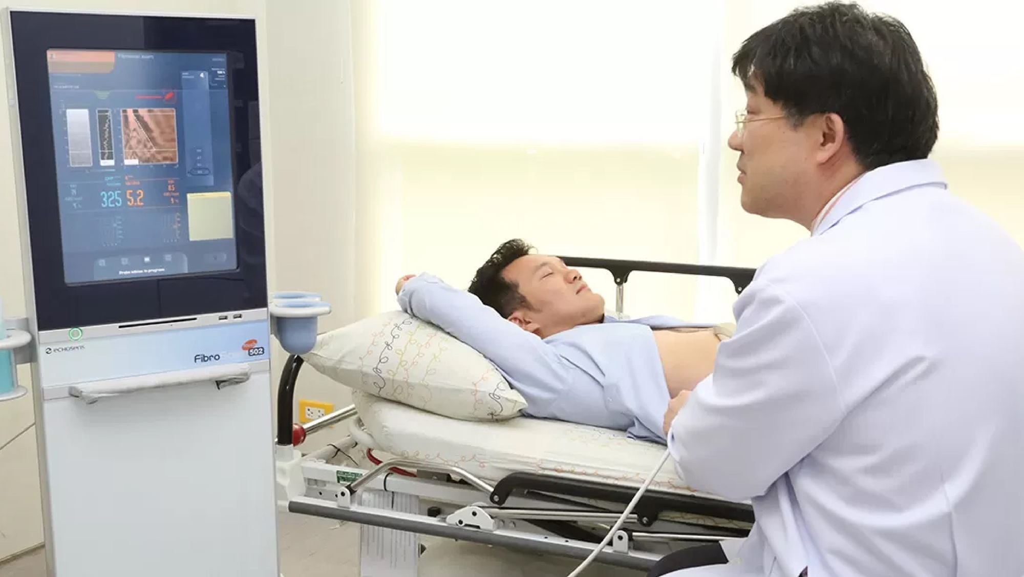
How Fibro Scan Works
- The patient lies on their back with the right arm raised behind the head.
- The technician identifies the appropriate intercostal space (usually over the right lobe of the liver).
- A water-based gel is applied to improve ultrasound conduction.
- A specialized probe (similar to an ultrasound probe) is placed against the skin.
- The device emits a mechanical pulse (vibration) that generates a low-frequency shear wave (~50 Hz) into the liver tissue.
- Simultaneously, the probe uses ultrasound pulses to detect the velocity of the shear wave. The faster it travels, the stiffer the liver tissue.
- The speed is converted into a stiffness value (kPa), reflecting fibrosis severity.
- The same probe measures ultrasound attenuation to assess hepatic steatosis. CAP values (dB/m) estimate liver fat content.
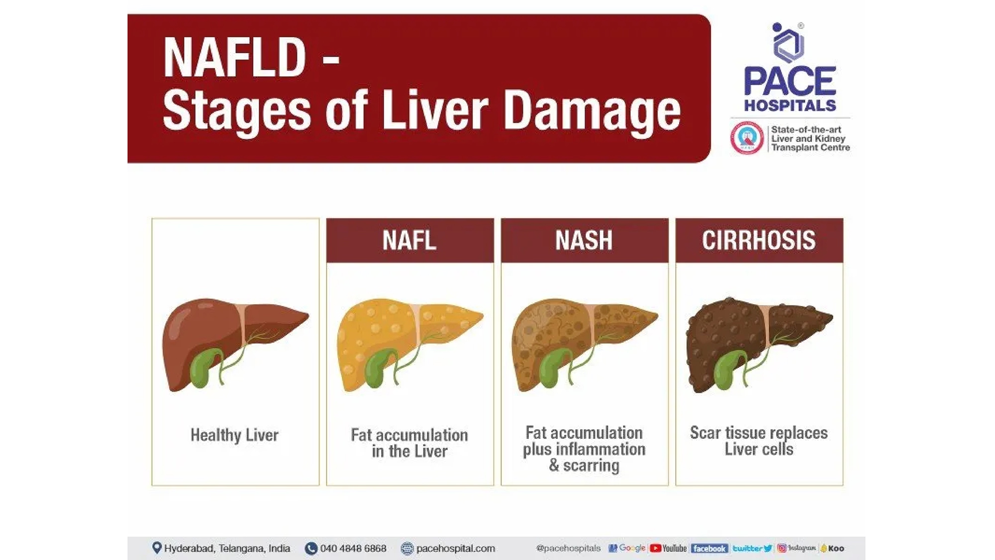
Key Benefits of Fibro Scan
- Accurate Quantification: Provides measurements of liver stiffness, fibrosis, and fat content (CAP score); detects and grades liver steatosis; stages liver damage (F0–F4, S0–S3).
- Early Detection and Prevention: Detects liver changes before symptoms appear; helps prevent progression to cirrhosis, liver failure, or hepatocellular carcinoma (HCC).
- Supports Personalized Treatment: Enables tailored management based on liver condition.
- Endorsed by major hepatology associations (EASL, AASLD) for assessing chronic hepatitis B/C, NAFLD/NASH, alcoholic liver disease, and metabolic syndrome-related liver issues.
12. CT Intra-Abdominal Fat
What is CT Intra-Abdominal Fat?
CT Intra-Abdominal Fat is a specialized type of CT imaging used to assess the amount and distribution of fat inside the abdominal cavity, particularly visceral fat surrounding internal organs such as the liver, pancreas, and intestines. This method is accurate and allows differentiation between visceral fat and subcutaneous fat.
How CT Intra-Abdominal Fat Works
- CT imaging (often low-dose, non-contrast) captures cross-sectional slices of the abdomen.
- Specialized software analyzes pixel densities to differentiate fat from muscle, bone, and organs.
- A quantitative report is generated showing total visceral fat volume, total subcutaneous fat volume, and visceral-to-subcutaneous fat ratio.
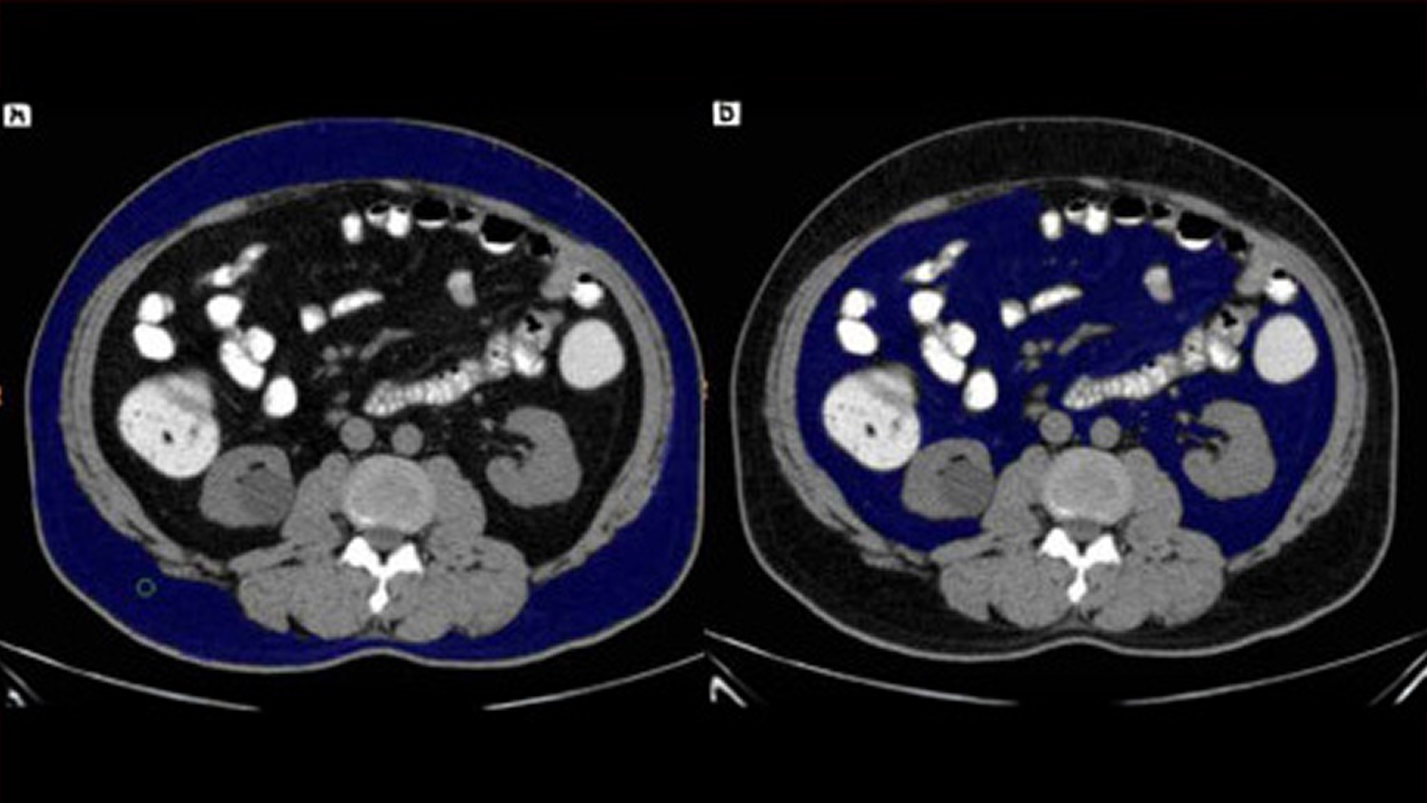
Key Benefits of CT Intra-Abdominal Fat
- Accurately quantifies visceral and subcutaneous fat.
- Assesses cardiometabolic risk beyond BMI or weight.
- Detects hidden fat even in normal-weight individuals.
- Guides personalized weight loss or metabolic therapies.
- Monitors progress from lifestyle or medical interventions.
- Supports early prevention of diabetes, heart disease, and fatty liver.
13. A Kidney Ultrasound
What is a Kidney Ultrasound?
A kidney ultrasound, also known as renal ultrasound, is a non-invasive imaging test that uses sound waves to create pictures of the kidneys and surrounding structures. It helps doctors assess size, shape, and position, and detect abnormalities like cysts, tumors, or blockages. It is safe, painless, and commonly used to diagnose and manage kidney-related conditions.
How a Kidney Ultrasound Works
- A technician applies gel to the skin over the kidneys (back or side).
- A transducer is moved over the skin, sending sound waves.
- The waves bounce off organs and tissues; echoes are collected.
- A computer converts echoes into images.
- The technician may ask for breath-holding or repositioning.
- The procedure takes about 30–60 minutes.
Key Benefits of a Kidney Ultrasound
- No needles, incisions, or contrast dyes.
- No radiation exposure.
- Quick and convenient with same-day results.
- Detects kidney stones, cysts, tumors, masses, or hydronephrosis.
- Doppler mode can show kidney blood flow.
- Helps guide treatment for abnormal kidney function or recurrent infections.
14. Thin Prep PAP Smear Female
What is Thin Prep PAP Smear Female?
A Thin Prep Pap Smear (liquid-based cytology) is a cervical cancer screening test using a liquid medium to collect cells for clearer, more accurate evaluation than conventional Pap smears. It detects abnormal, precancerous, or cancerous cells in the cervix.

How Thin Prep PAP Smear Female Works
- A provider gently collects cervical cells using a brush or spatula.
- The vial is processed to remove mucus, blood, and debris.
- A thin, even layer of cells is placed on a slide.
- A cytopathologist examines the slide for abnormalities.
Key Benefits of Thin Prep PAP Smear Female
- More accurate detection of abnormal or precancerous cells.
- Helps prevent cervical cancer through early identification.
- Allows combined Pap + HPV testing from the same sample.
- Quick, simple, and minimally uncomfortable.
15. Mammogram Breast Female
What is Mammogram Breast Female?
A mammogram is a specialized low-dose X-ray of the breast used to screen for and detect breast cancer and abnormalities.
- Screening mammogram: for women without symptoms.
- Diagnostic mammogram: for symptoms like lumps, discharge, or pain.
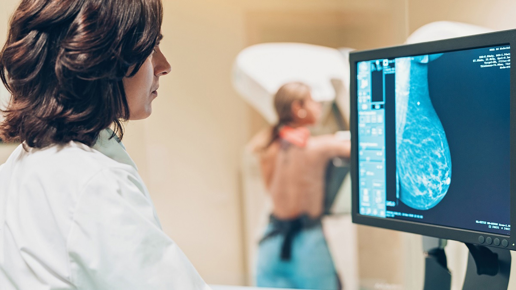
How Mammogram Breast Female Works
- Breast is positioned on a detector plate.
- Gentle compression spreads tissue for clearer imaging and less radiation.
- Low-dose X-rays capture images from several angles.
- A radiologist reviews images for masses, calcifications, or distortions.
Key Benefits of Mammogram Breast Female
- Life-saving early cancer detection.
- Reduces need for aggressive treatments.
- Creates a personal baseline for future comparison.
- Identifies malignant, benign, and pre-cancerous changes.
16. U/S Breast Female
What is U/S Breast Female?
A breast ultrasound uses sound waves to create images of breast tissue. It is safe, painless, radiation-free, and used to evaluate lumps, distinguish cysts from tumors, and guide biopsies.
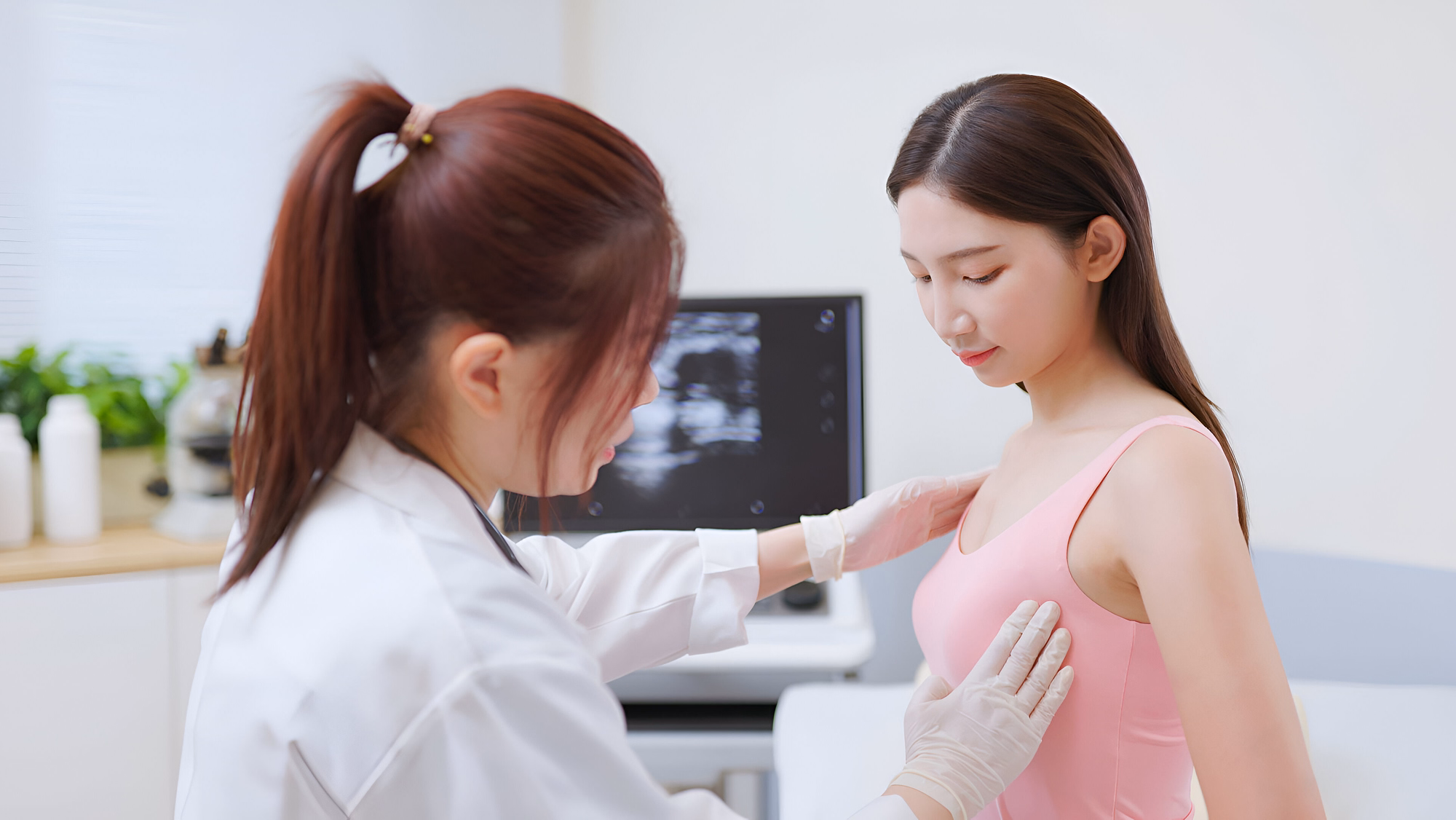
How U/S Breast Female Works
- A transducer is placed on the breast.
- Gel improves sound conduction.
- High-frequency sound waves generate echoes.
- A computer converts echoes into real-time images.
- A radiologist interprets abnormalities.
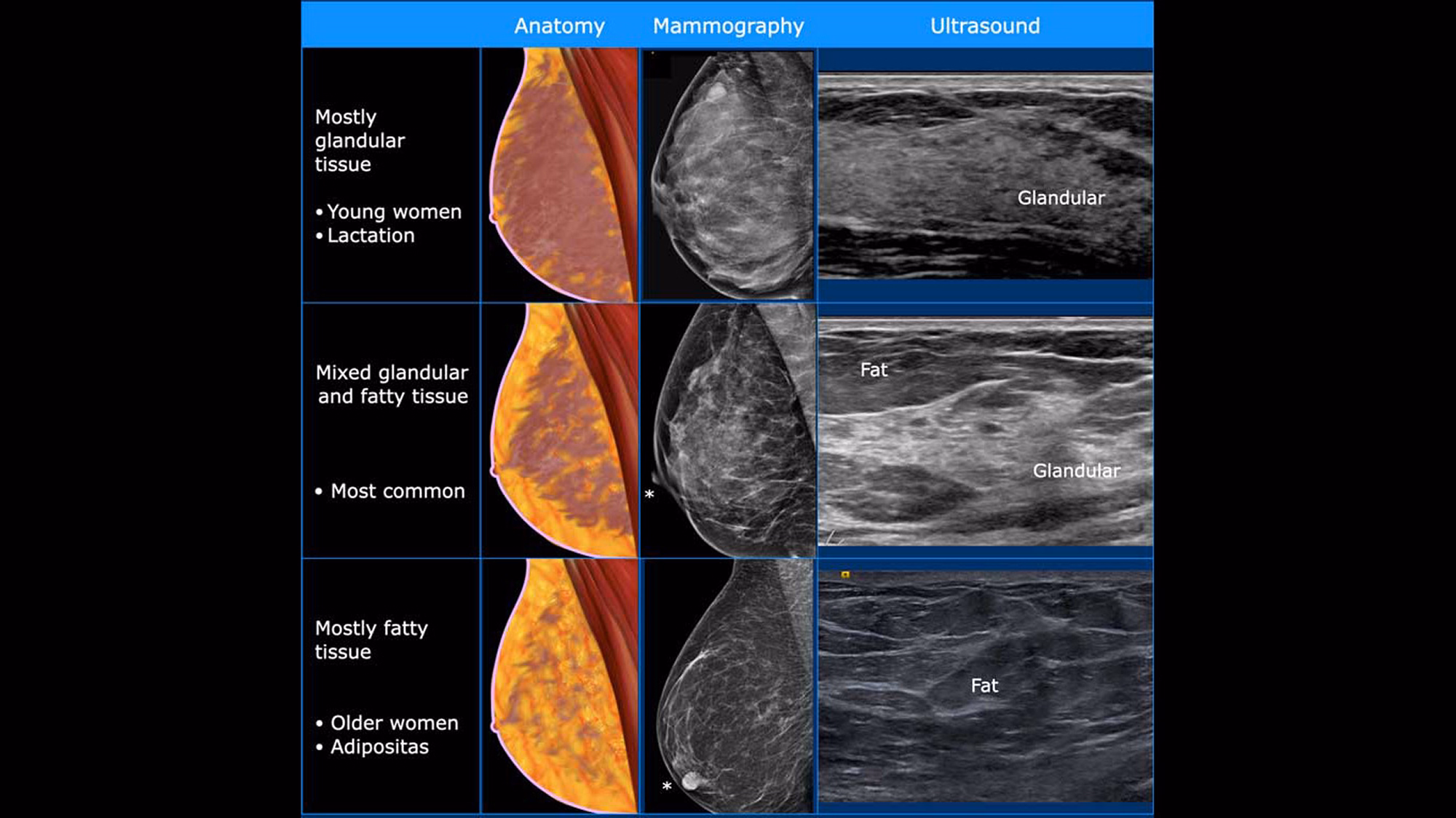
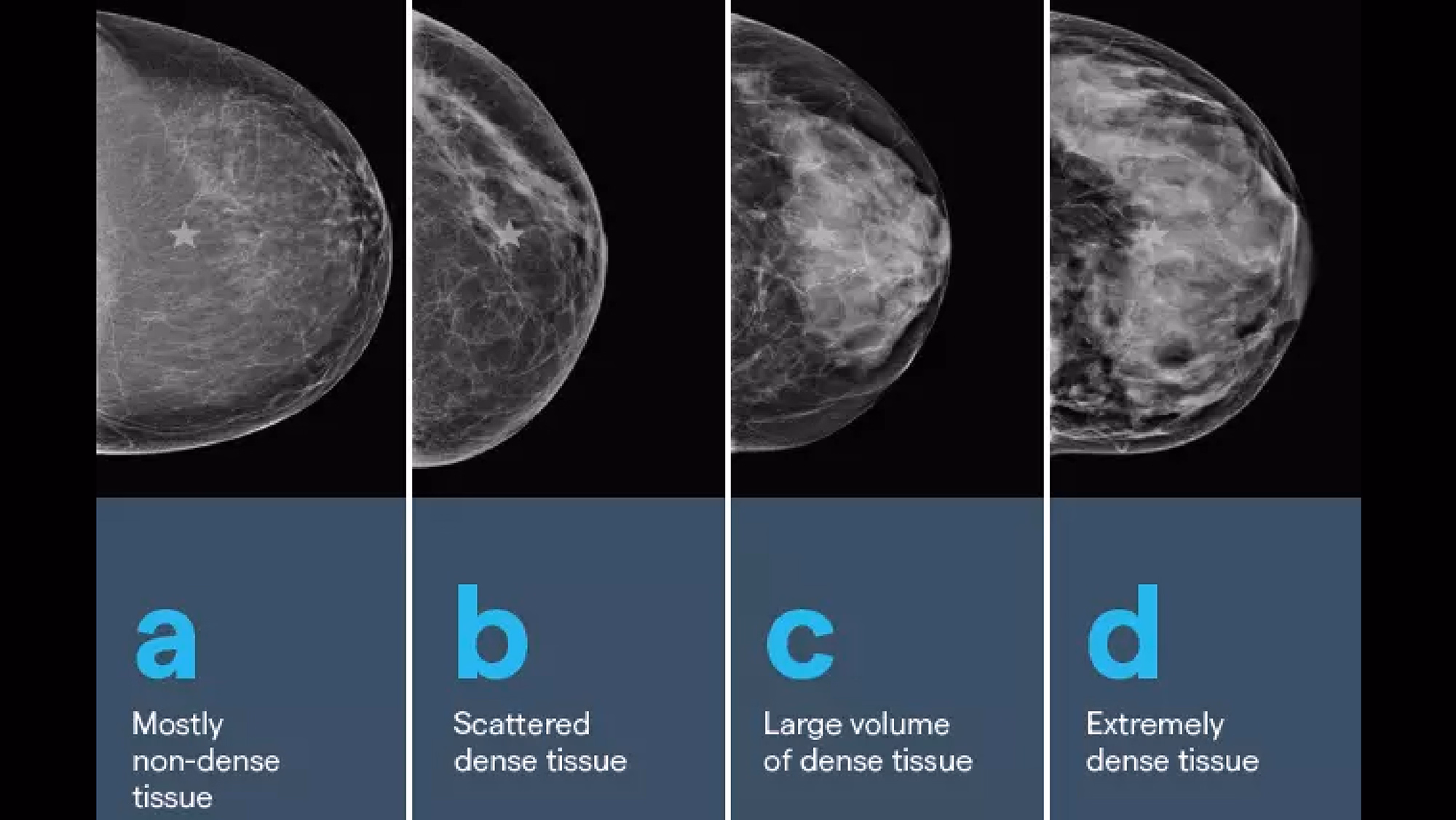
Key Benefits of U/S Breast Female
- Effective in dense breast tissue.
- Differentiates solid vs. fluid masses.
- Fast procedure (15–30 minutes).
- Complements mammography and MRI.
Total Body Health Checkup is more than a routine examination. It offers a comprehensive solution to assess internal balance, restore organ vitality, and promote long-term wellness throughout the body.
As part of an integrated Wellness & Longevity Program, it provides the foundation for personalized health optimization, empowering you to prevent disease, restore balance, and prepare for advanced therapies such as Stem Cell, EBOO, and DFPP. It is not merely a checkup, but a strategic investment in long-term vitality and wellness from within.
Preparation Guidelines
- Drink a small amount of water for smoother blood collection.
- Get 6–8 hours of sleep and avoid stress or heavy exercise.
- Inform your doctor if you have chronic conditions or take regular medication.
- Female clients should avoid testing during menstruation, especially breast or cervical exams.
Recovery Care
- Rest and eat a healthy meal to recover energy.
- Drink plenty of water, especially after CT or MRI with contrast.
- Avoid strenuous activity on the same day.
- Review results with your doctor to plan further wellness or treatment programs.